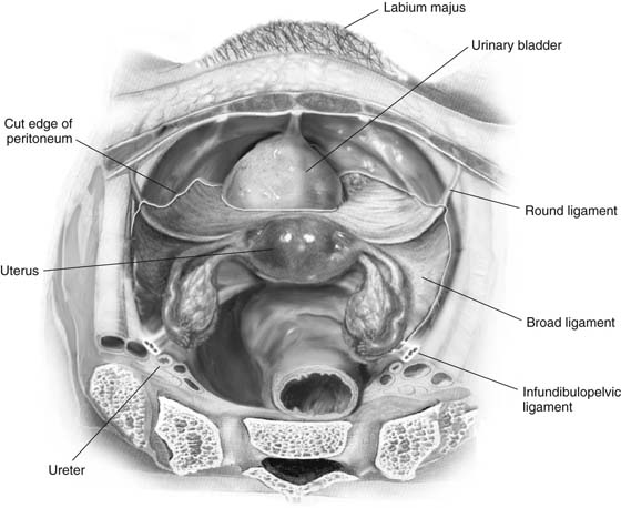
The uterosacral ligament connects the uterus at the level of thecervix to the sacrum and is therefore its primary support. Introduction to pelvic anatomy 1. 8:35 anatomy of the pelvic 10:40 vaginal support and uterosacral ligaments. There are many organs that sit in the pelvis, including much of the urinary system, and lots of the male or female reproductive systems. Pelvic floor anatomy & function:
Pelvis And Hip Joint Amboss from media-us.amboss.com The pelvic girdle consists of two symmetrical halves. With inks to related posts. Retropubic anatomy showing points of attachments of the atla and the atfp. Functional anatomy of the male pelvic floor online course: • muscles and ligaments form a pelvic floor. Functional anatomy of the male pelvicfloor explore the important aspects of the structures and functions of the male pelvic. Published on 09/03/2015 by admin. Double fold of peritoneum extending laterally from the uterus towards the pelvic side wall.
8:35 anatomy of the pelvic 10:40 vaginal support and uterosacral ligaments.
8:10 pelvic sidewall anatomy and retroperitoneal spaces. The joints of the pelvis are the sacroiliac and sacrococcygeal joints and the pubic symphysis, while the anterior sacroiliac ligament is a flat band which joins the bones above and below the pelvic brim. Amis, a and g dawkins. Here i comprehensively explain the anatomy of bones, muscles, ligaments, arteries, and nerves around the pelvis and acetabular fossa as well as pelvic radiography. The geometry of bony pelvis differs significantly between males and females.
Intra Abdominal Pelvic Anatomy Obgyn Key from obgynkey.com This chapter will focus on those aspects of pelvic anatomy that have special importance to the practice of obstetrics. 8:10 pelvic sidewall anatomy and retroperitoneal spaces. This mri male pelvis axial cross sectional anatomy tool is absolutely free to use. Published on 09/03/2015 by admin. Related online courses on physioplus. Various pelvic ligaments help support the uterus and other pelvic organs. 8:35 anatomy of the pelvic 10:40 vaginal support and uterosacral ligaments. Introduction to pelvic anatomy 1.
Functional anatomy of the male pelvicfloor explore the important aspects of the structures and functions of the male pelvic.
Functional anatomy of the anterior cruciate ligament. Related online courses on physioplus. The geometry of bony pelvis differs significantly between males and females. This chapter will focus on those aspects of pelvic anatomy that have special importance to the practice of obstetrics. • pelvis begins at the iliac crests and ends at the symphysis pubis.
Anatomy Of The Pelvic Girdle Physiopedia from www.physio-pedia.com Uterus location and anatomical relations. Various pelvic ligaments help support the uterus and other pelvic organs. Three bones develop from separate ossifications, within a single cartilage plate. Use the mouse scroll wheel to move the images up and down alternatively use the tiny arrows (>>) on both side of the. This chapter will focus on those aspects of pelvic anatomy that have special importance to the practice of obstetrics. Functional anatomy of the anterior cruciate ligament. Introduction to pelvic anatomy 1. The bony pelvis & gender differences in pelvic anatomy.
The pelvic girdle consists of two symmetrical halves.
There are many organs that sit in the pelvis, including much of the urinary system, and lots of the male or female reproductive systems. As a result, all who perform surgery in the chapter 2 abdominal and pelvic anatomy 11. This mri male pelvis axial cross sectional anatomy tool is absolutely free to use. 494 raizada & mittal the uterosacral ligaments extend from the upper portion of the cervix posteriorly to the third sacral. Related online courses on physioplus. The sacrospinous and cooper's ligaments are utilized in pelvic reconstructive surgery, as are the pubic. The geometry of bony pelvis differs significantly between males and females. 8:10 pelvic sidewall anatomy and retroperitoneal spaces. Pelvic skeleton includes two hip bones, sacrum and coccyx. The pelvis (plural pelves or pelvises) is either the lower part of the trunk of the human body between the abdomen and the thighs (sometimes also called pelvic region of the trunk) or the skeleton embedded in it (sometimes also called bony pelvis, or pelvic skeleton). Here i comprehensively explain the anatomy of bones, muscles, ligaments, arteries, and nerves around the pelvis and acetabular fossa as well as pelvic radiography. 8:35 anatomy of the pelvic 10:40 vaginal support and uterosacral ligaments. ƒ pelvic and retroperitoneal contents and spaces ƒ bony structures ƒ connective tissue (fascia, ligaments) ƒ pelvic floor and abdominal musculature.
Uterus location and anatomical relations pelvic anatomy. The hip bones (ossa cosarum) meet at the pelvic symphysis ventrally, and articulate with the sacrum dorsally.
